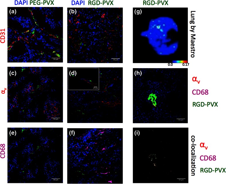
Figure 5.
Immunofluorescence analysis of tumor and lung sections. The tumor sections were stained with antibodies specific for the endothelial marker CD31 (red, a + b), integrin α v [red, c + d (inset: higher magnification)], the tumor macrophage marker CD68 (pink, e + f) and DAPI (blue) to determine the localization of PEG-PVX and RGD-PVX within the tumor. (g–i) Whole lung (g) and lung sections (h, i) from mice treated with RGD-PVX were stained for α v integrin (red) and CD68+ macrophages (pink). Co-localization analysis was used to highlight the hotspots of RGD-PVX accumulation with integrins and CD68 macrophages (i). Scale bars = 50 μm.

