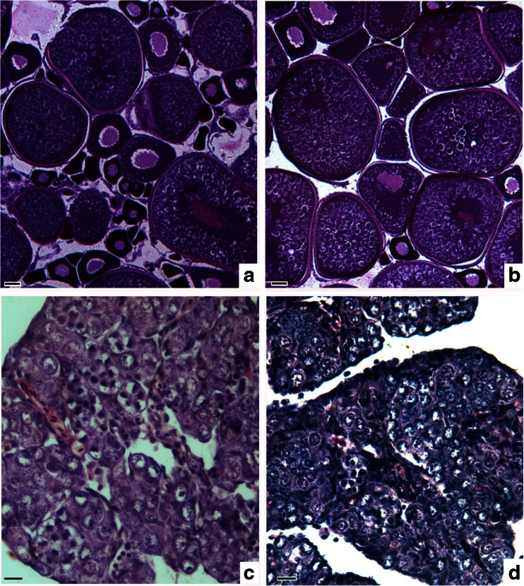
Fig. 5.
Gonadal microstructure in RCC, 4nF1, and G1. a The ovary of RCC, showing normally developed oocytes in phase III; b The ovary of G1, showing normally developed oocytes in phase III; c The ovary of 4nF1 is occupied by many oogonia; d Abnormal ovary of G1, showing proliferating oogonia that did not develop into oocytes. Bar in a–d, 50 μm

