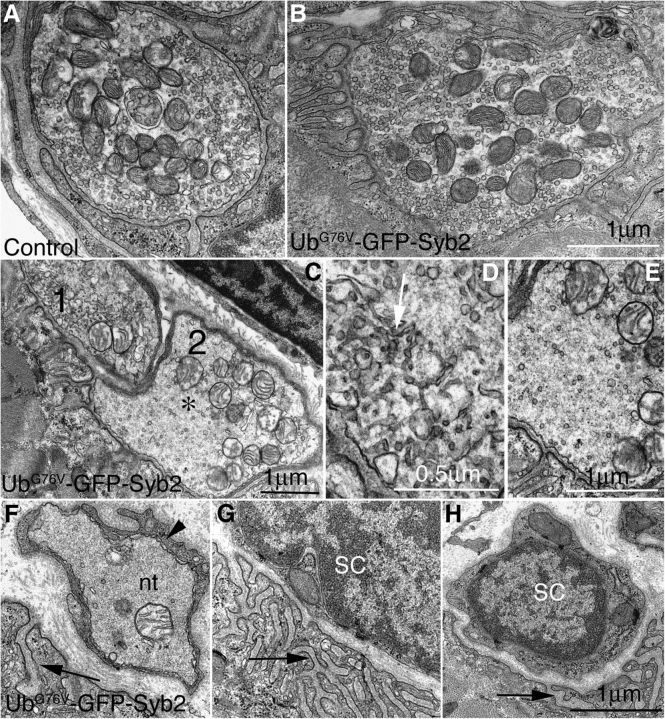
Figure 13.
Ultrastructural defects of motor nerve terminals in UbG76V-GFP-Syb2 mice (8 months of age). A, B, Examples of normal NMJs in control mice (A) and UbG76V-GFP-Syb2 mice (B). C, An example of an abnormal NMJ from UbG76V-GFP-Syb2 mice. This NMJ contains two nerve terminals (marked by 1 and 2), which are further magnified in D and E. The density of synaptic vesicles is markedly reduced; the axoplasm appears amorphous [denoted by an asterisk (*)] and is characterized by the accumulation of abnormal tubulovesicular structures (D, white arrow). F–H, Examples of degenerated NMJs: a nerve terminal engulfed by a Schwann cell process (F, black arrowhead), and synapses devoid of motor nerve terminals (G, H). Synaptic sites are marked by the presence of postsynaptic junctional folds (F–H, black arrow). nt, Nerve terminal; SC, Schwann cell. Scale bars: A–C, E–H, 1 μm; D, 0.5 μm.

