Figure 1.
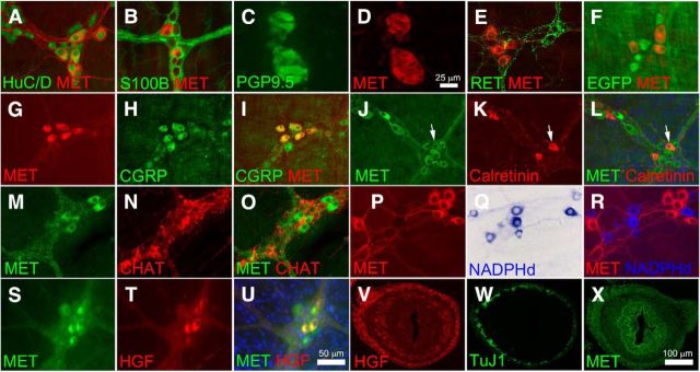
MET immunoreactivity was detected in a subset of myenteric plexus CGRP+ IPANs. A, MET immunoreactivity was detected in 34 ± 6% of HuC/D+ myenteric neurons of the adult mouse small bowel. All MET+ cells expressed the pan-neuronal marker HuC/D. B, MET staining was absent from S100B+ enteric glia. C, D, MET was detected in human myenteric neurons using colon cross sections. E, F, MET and RET are detected in mutually exclusive sets of neurons as confirmed by immunohistochemistry (E) and by using a RET-EGFP reporter mouse (F). G–I, 100% of MET+ neurons were CGRP+ and 50% of CGRP+ neurons were MET+. J–L, MET was also found in 16 ± 5% of calretinin+ cells and 8 ± 2% of MET+ cells are calretinin+. Arrows highlight a MET+ calretinin+ neuron. M–O, 12 ± 0.1% of ChAT+ cells were MET+ and 8 ± 0.1% of MET+ neurons were ChAT+. P–R, There was no overlap between MET and NADPH diaphorase-stained nitric oxide-producing neurons. S–U, HGF was detected in 43 ± 11% of MET+ neurons and 100% of the HGF+ neurons were MET+. V, In cross sections of E14.5 fetal bowel, HGF-IR was found in mesenchymal cells surrounding developing ENS stained with TuJ1 (W). X, At E14.5, MET+ cells were present in the region of developing ENS as well as in gut epithelium and mesenchymal cells. Scale bars in U applies to A, B, E–U. Scale bar in D applies to C and D. Scale bar in X applies to V–X. N ≥ 3 replicates/staining condition.

