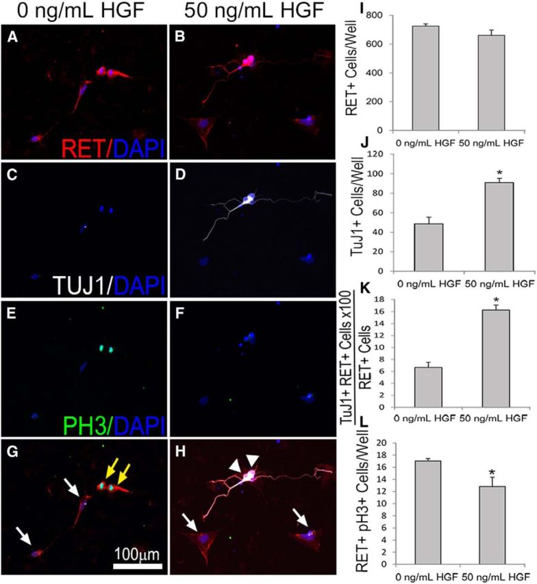
Figure 3.

HGF/MET signaling enhanced ENS precursor differentiation into neurons in vitro. A–H, E12.5 ENS precursors were maintained in culture after p75NTR immunoselection for 2 d in the presence or absence of 50 ng/ml HGF before immunohistochemistry using RET (A, B), TuJ1 (C, D), and pH3 (E, F) antibodies as well as DAPI nuclear staining (A–H). G, H, Merged images. I–K, While the total number of RET+ cells was not altered by HGF (I), the total number of TuJ1+ neurons (J) and the percentage of RET+ cells that were TuJ1+ (K) increased with HGF treatment. L, The number of dividing precursor cells (pH3 and RET double positive) decreased with HGF treatment, suggesting that HGF increased neuronal differentiation and decreased proliferation. White arrows, Nonmitotic RET+ pH3− ENS precursors. Yellow arrow, Mitotic RET+PH3+ ENS precursors. White arrowhead, RET+TuJ1+ neurons. Scale bar in G applies to all images (N = 3 biological replicates/group; 12 individual wells/group; *p < 0.01, Student's t test).
