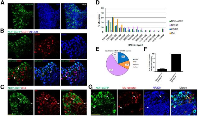
Figure 6.
Various types of DRG neurons express NOP-eGFP receptors. To characterize the NOP-eGFP distribution in DRG neurons, sections were incubated with anti-GFP antibody together with anti-μ receptor antibody, DRG markers; anti-CGRP, -NF200 antibodies, or biotinylated IB4. A, NOP-eGFP expression in DRG neurons (green). Nuclei were stained with DAPI (blue). The NOP-eGFP containing DRG neurons were quantified by determining the percentage of eGFP-positive cells compared with the total number of sensory neurons (n = 1396). The total number of DRG sensory neurons was determined by counting the total number of DAPI-stained cells and excluding those from glia and connective tissues. Tissue sections were also costained with anti-CGRP and -NF200 antibodies, (B) in which the white arrowheads indicate NOP-eGFP+, CGRP-myelinated medium DRG neurons, or (C) biotinylated IB4. In each panel, white arrows indicate the cells where costaining occurs. D, Size profiling of DRG neurons that are expressing NOP-eGFP, CGRP, or NF200, or bind to IB4. E, Identity of NOP-eGFP+ DRG neurons. F, Percentage of medium NOP-eGFP+ DRG neurons that are myelinated and not coexpressing CGRP. Data are represented as mean ± SEM, G, colocalization of NOP-eGFP and μ receptors in DRG. Small unmyelinated neurons coexpress NOP-eGFP and μ receptors. White arrows depict the cells coexpressing NOP-eGFP and μ receptors. Scale bars, 100 μm.

