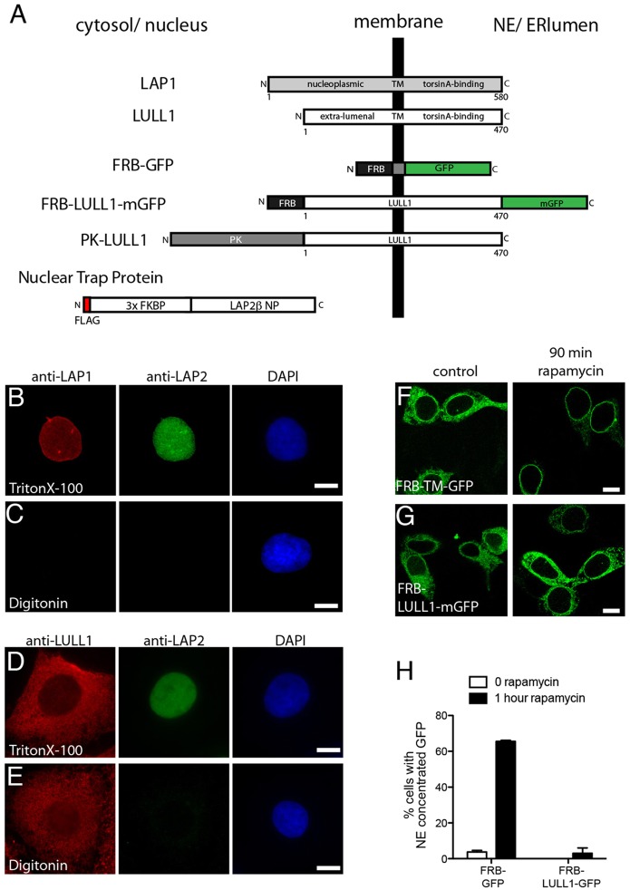
Fig. 1.
LAP1 and LULL1 have distinct localization in the ER system. (A) Schematic showing protein organization. (B,C) Differential permeabilization studies place LAP1 on the INM. An antibody recognizing the LAP1 nucleoplasmic domain detects nuclear-envelope-concentrated LAP1 in U2OS cells permeabilized with Triton X-100 but not digitonin, as is also observed for the control nuclear LAP2 protein. (D,E) Differential permeabilization studies place LULL1 in peripheral ER and ONM. An antibody recognizing the LULL1 cytoplasmic domain detects LULL1 throughout the cell following permeabilization with either Triton X-100 or digitonin. Nuclear LAP2 is only detected in Triton X-100 sample. (F,G) Rapamycin trapping demonstrates accumulation of control FRB–TM–GFP but not FRB–LULL1–mGFP in the INM. Images show (F) FRB–TM–GFP and (G) FRB–LULL1–mGFP in NIH-3T3 cells before (left panels) or after 90 min incubation (right panels) with rapamycin. Scale bars: 10 µm. (H) Quantification of the percentage of cells with nuclear-envelope-concentrated GFP (mean±s.e.m.). Data are from three transfections, each counting >50 cells.

