Fig. 4.
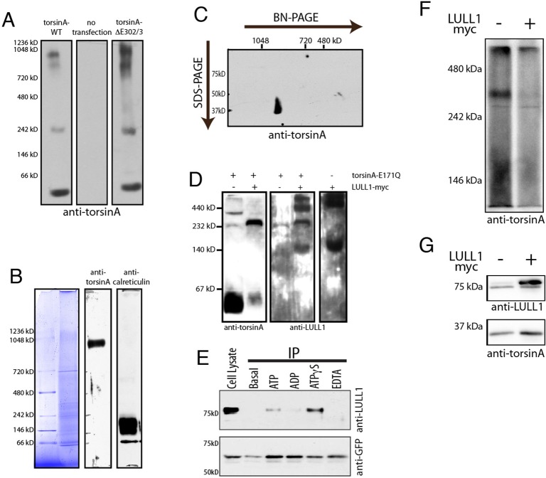
Multimeric torsinA complexes are sensitive to LULL1 and ATPase activity. (A) Wild-type and torsinA-ΔE302/3 seen in high molecular mass structures by BN-PAGE. Panels show immunoblotting of 20 µg of 0.5% digitonin-solubilized cell lysate from NIH-3T3 cells transfected as indicated. (B) A high molecular mass torsinA species in mouse liver microsomes. 20 µg of 1% digitonin-solubilized microsomal protein separated on a 3.2–16% gradient BN-PAGE, with individual lanes stained for total protein or immunostained for torsinA and calreticulin. Anti-torsinA immunoreactivity is seen at ∼900 kDa. (C) Anti-torsinA immunoblotting following second dimension SDS-PAGE confirms that the 37 kDa torsinA is present in the high molecular mass immunoreactive species. (D) TorsinA-E171Q and LULL1 co-assemble into an ∼250 kDa species consistent with formation of a stable hetero-hexameric complex. Panels show anti-torsinA and anti-LULL1 immunoblotting of BN-PAGE separated lysates from U2OS cells transfected with torsinA-E171Q, LULL1–Myc, or both. Co-expressed torsinA-E171Q and LULL1 are present in an ∼250 kDa band that is absent when either one is overexpressed alone. (E) ATP hydrolysis destabilizes the LULL1–torsinA complex monitored by co-immunoprecipitation (IP) from NIH-3T3 cells stably expressing GFP–torsinA. Upper panel, lane one, shows total cellular LULL1 (Cell Lysate). Lanes 2–6 show the amount of LULL1 that co-immunoprecipitates with GFP–torsinA without added nucleotides, or upon the addition of 2 mM ATP, ADP, ATPγS or EDTA. The lower panel shows anti-GFP immunoblotting, which confirms immunoprecipitation of similar amounts of GFP–torsinA. (F) BN-PAGE shows that LULL1 expression decreases the ∼250–300 kDa species of torsinA resolved by BN-PAGE of digitonin-solubilized samples. Shown is anti-torsinA immunoblotting of BN-PAGE-separated lysates from U2OS cells transfected with wild-type torsinA alone or together with LULL1–Myc and solubilized in 1% digitonin. BN-PAGE separation is on a 7.5% gel. This experiment was repeated three times with the same result. (G) SDS-PAGE and immunoblotting shows torsinA and LULL1 in the samples resolved by BN-PAGE in F.

