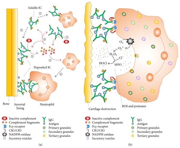
Figure 1.
Interaction among neutrophils, immune complexes, and the complement system in mediating joint damage in rheumatoid arthritis. (a) Soluble ICs in the rheumatoid synovial fluid can bind and stimulate primed neutrophils (①) or deposit in the synovial lining (②). Both soluble and deposited ICs activate the complement system (③), generating protein fragments; these components of the activated complement system deposit in synovial tissues, opsonize soluble and tissue-bound ICs (④), and attract neutrophils to the inflamed joint (⑤). The recruited neutrophils recognize IgG and some complement fragments contained in the ICs via their Fcγ and complement receptors, respectively (⑥). (b) Tissue-bound ICs activate neutrophils, which are not able to phagocytose them; as a consequence, neutrophils release ROS and proteolytic enzymes from their granules and secretory vesicles to the extracellular milieu. This process is termed “frustrated phagocytosis.” The cytotoxic products can overwhelm the local antiprotease and antioxidant protective mechanisms and degrade components of articular cartilage. CR1/CR3, complement receptors types 1 and 3; IC, immune complex; IgG, immunoglobulin G; MPO, myeloperoxidase; ROS, reactive oxygen species. This illustration was adapted from the review article published by Wright et al [21], with permission of Macmillan Publishers Limited.

