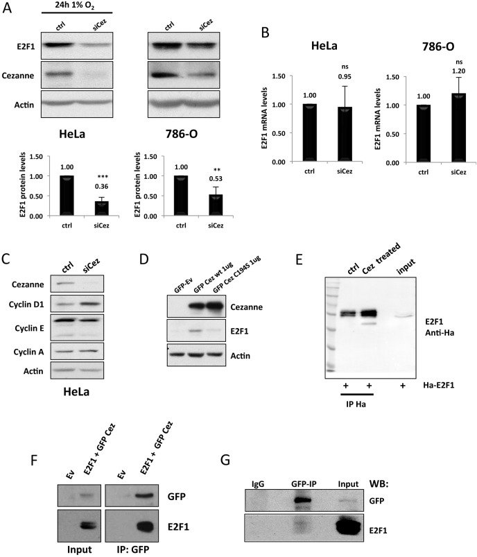
Fig. 4.
Cezanne regulates E2F1 protein stability. (A) HeLa and 786-O cells were transfected with either a non-targeting control (ctrl) or a Cezanne-targeting siRNA (siCez). At 24 h after transfection, HeLa cells were exposed to 1% O2, and whole cell lysates of both HeLa and 786-O cells were prepared after a further 24-h incubation. Whole cell extracts were analysed by western blotting to assess total levels of E2F1 and Cezanne. The band intensities were measured, and the values were normalised to those in cells transfected with the control siRNA (P-values are significant according to the Student's t-test; ***P<0.001). The fold-changes relative to the control are shown above the bars. (B) HeLa and 786-O cells were treated as in A, but the total RNA was extracted, and the transcript levels of E2F1 were analysed by using RT-qPCR. ns, not significant. (C) Whole cell extracts from HeLa cells that had been treated as described in A were analysed by western blotting with the antibodies indicated. (D) HeLa cells were transfected with either a wild-type (wt) or catalytically inactive (C194S) GFP–Cezanne construct, or with empty vector (Ev), and whole cell lysates were analysed by western blotting for the total E2F1 protein levels 48 h post transfection. (E) HEK293 cells were transfected with 5 µg of a HA–E2F1 construct for 48 h before lysis. Following immunoprecipitation (IP) with HA-beads, samples were treated where indicated with recombinant Cezanne (Cez). Lysates were analysed by western blotting with the indicated antibodies. (F) HEK293 cells were co-transfected with 5 µg of wild-type GFP–Cezanne and HA–E2F1 plasmids for 48 h before lysis. GFP–Cezanne was immunoprecipitated with an anti-GFP antibody, and lysates were analysed by western blotting with the indicated antibodies. (G) HEK293 cells were transfected with 5 µg of plasmid encoding wild-type GFP–Cezanne and left to express for 48 h. Cells were then lysed in a mild NP-40 lysis buffer, and GFP–Cezanne was immunoprecipitated with an anti-GFP antibody. Co-immunoprecipitation of endogenous E2F1 was analysed by western blotting (WB) as indicated.

