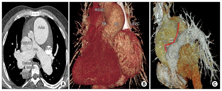
Fig. 1.
(A, B) Preoperative computed tomography. This image shows left isomerism, a dilated right superior vena cava, a dilated azygos vein, and pulmonary stenosis. The main PA is located to the right and posterior to the AAo. (C) Postoperative computed tomography. Despite the ascending aortic reduction plasty, the recipient distal AAo still push out into the main PA (Solid red line). Little space was available to insert the LSVC into the retro-aortic area. AAo, ascending aorta; RSVC, right superior vena cava; PA, pulmonary artery; LSVC, left superior vena cava; Ao, aorta.

