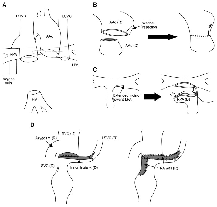
Fig. 2.
(A) Schematic drawing after the recipient’s heart excision. (B) V-shape reduction plasty of the recipient aorta. (C) The incision was extended toward the recipient’s LPA. The donor main pulmonary artery was transected at the distal part of the right pulmonary artery and then anastomosed to the recipient pulmonary artery. (D) Anastomosis between the donor SVC and innominate vein and the recipient right SVC and left SVC. SVC, superior vena cava; RSVC, right superior vena cava; AAo, ascending aorta; LSVC, left superior vena cava; RPA, right pulmonary artery; LPA, left pulmonary artery; HV, hepatic vein; R, recipient; D, donor.

