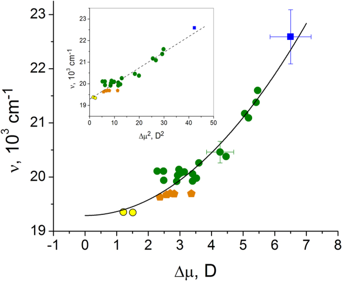
Figure 2. Dependence of the 0–0 transition frequency on the change of the permanent dipole moment for a GFP anionic chromophore in a number of different local environments.

The blue square represents the model HBDI− chromophore in alkaline D2O solution. Standard deviations are shown by bars. Twenty six different FP variants under investigation are sub-divided into three different groups, according to the local structure of their chromophore surrounding: (1) The mutants derived from EGFP and mTFP, where the two chromophore oxygen atoms, i.e. at phenolate and imidazolinone rings, maintain five hydrogen bonds with the surrounding, similar to the original EGFP and mTFP63,64,65,66, are shown by green circles; (2) EGFP-type of mutants containing, among others, the T203I mutation, which causes a reduction of the number of hydrogen bonds from five to four in the chromophore cluster, are shown by orange pentagons; (3) Yellow FP derivatives of GFP containing, among others, the T203Y mutation, which along with the reduction of the number of hydrogen bonds also causes a π-stacking interaction of the chromophore with the Tyr203 phenol38, are shown by yellow circles. The inset shows the same data in the linearizing, ν versus Δμ2 coordinates.
