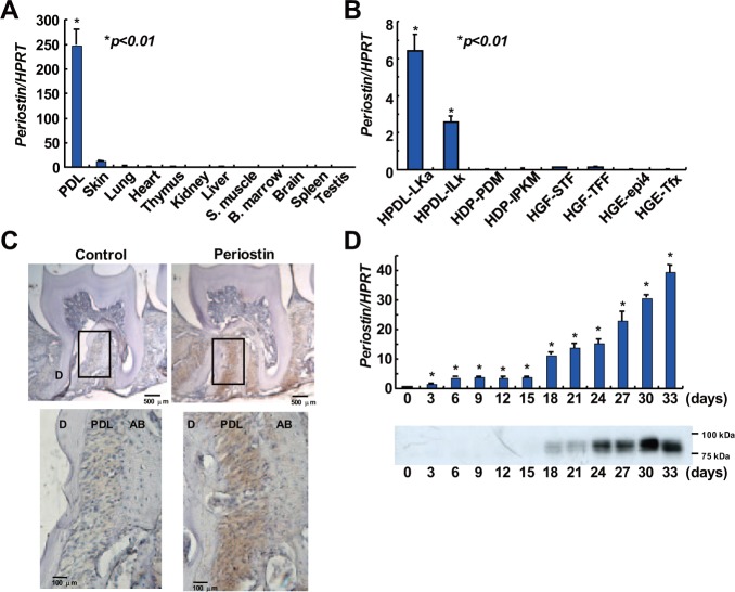
Figure 1.
Periostin is specifically expressed in periodontal ligament tissue and cells and is induced during PDL cell differentiation. Real-time PCR analysis of periostin in various human tissues (A) and human oral-tissue-derived cell lines (B). Values represent the mean ± SD of triplicate assays. *p < .01, compared with other tissues. **p < .01, compared with other cell lines. (C) Specific expression of periostin in PDL tissues in vivo. Periostin protein was detected in 8-week-old mouse periodontium by immunohistochemical staining with an anti-periostin antibody. D, dentin; PDL, periodontal ligament; AB, alveolar bone. (D) Induction of periostin mRNA and protein during PDL cell differentiation. Human PDL cells were cultured in mineralization-inducing medium. Periostin expression was assessed by real-time PCR analysis (upper panel) and Western blot analysis (lower panel) on every third day of culture. Values represent the mean ± SD of triplicate assays. *p < .01, compared with day 0.

