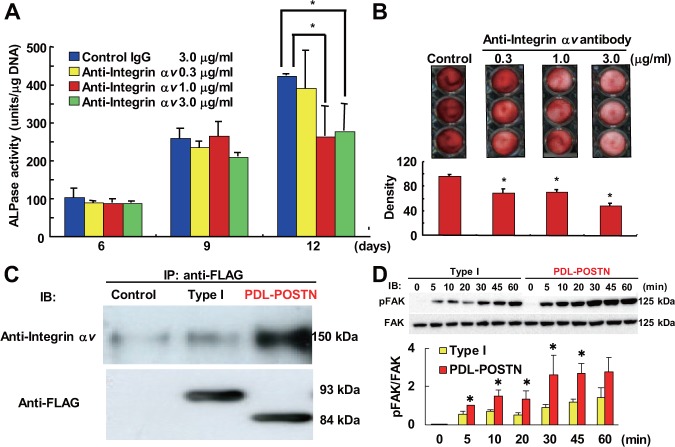
Figure 4.
PDL-POSTN positively regulates PDL cell differentiation through a direct interaction with integrin αvβ3. (A) MPDL22 cells transfected with PDL-POSTN were cultured in mineralization-inducing medium in the presence of a neutralizing antibody against integrin αv. ALPase activity of these cells was measured on the indicated day of culture. Values are the mean ± SD of triplicate assays. (B) Cells prepared the same way as in (A) were treated with an anti-integrin αv neutralizing antibody to investigate its effect on calcified nodule formation induced by PDL-POSTN. Alizarin red staining was performed on day 12. The image illustrates alizarin red staining, while the chart below shows the quantification of this staining. Values are the mean ± SD of triplicate assays. (C) FLAG-tagged recombinant periostin was incubated with recombinant integrin αvβ3 and immunoprecipitated with an anti-FLAG antibody. Co-precipitated recombinant integrin αvβ3 was detected with an anti-integrin αv antibody (upper panel). Input recombinant periostin was detected with anti-FLAG antibody (lower panel). (D) MPDL22 cells were stimulated with immobilized Type I periostin or PDL-POSTN at the indicated times, and phosphorylated FAK (pFAK) was detected by Western blot analysis. Quantitative analysis is shown as the ratios of phosphorylated-FAK to total FAK, determined by densitometric analysis. Values represent the mean ± SD of three independent assays. *p < .05 compared with Type I periostin. Control, conditioned medium derived from MPDL22 cells infected with a LacZ-expressing adenovirus; Type I, conditioned medium derived from MPDL22 cells infected with an adenovirus expressing the periostin type I isoform; PDL-POSTN, conditioned medium derived from cells infected with an adenovirus expressing PDL-POSTN. This figure is available in color online at http://jdr.sagepub.com.

