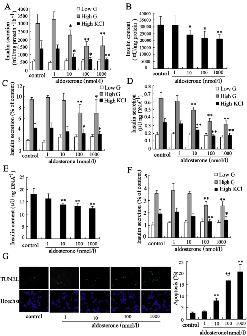
Figure 1. Aldosterone induced dysfunction and apoptosis of clonal β-cell.
Treatment with aldosterone (10, 100, 1000 nmol/l) for 24 h significantly decreased (A) insulin secretion (white bars, 2 mmol/l glucose; gray bars, 20 mmol/l glucose; black bars, 50 mmol/l KCl), (B) insulin content and (C) ratio of secreted insulin to insulin content represented as percentage of Min6 cells. Similar effects on (D) insulin secretion (white bars, 2 mmol/l glucose; gray bars, 20 mmol/l glucose; black bars, 50 mmol/l KCl), (E) insulin content of mouse islets and (F) ratio of secreted insulin to insulin content represented as percentage of mouse islets were observed. (G) Treatment with aldosterone (10, 100, 1000 nmol/l) for 72 h significantly induced apoptosis of Min6 cells measured by staining with TUNEL and Hoechst. Apoptosis was determined by scoring the percentage of TUNEL-positive cells. About 2,000 cells were scored for each group in one experiment. *P < 0.05 and **P < 0.01, compared to control.

