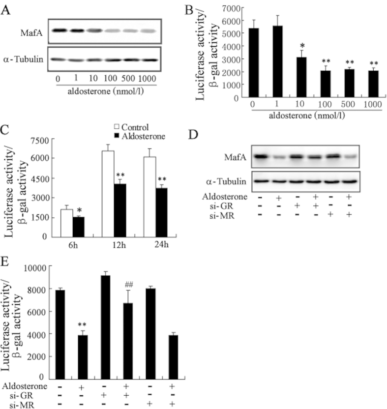
Figure 3. Aldosterone treatment results in a decrease in MafA protein level and transcriptional activity in GR-dependent pathway.
(A) Treatment with aldosterone in Min6 cells for 24 h significantly decreased the protein levels of MafA in a dose-dependent manner. (B) Min6 cells were transfected with MAREs-luc reporter plasmid for 24 h, then, treated with aldosterone for 24 h. Treatment with aldosterone significantly decreased MafA transcriptional activity in a dose-dependent manner. (C) Min6 cells were transfected with MAREs-luc reporter plasmid for 24 h, then, treated with aldosterone for 6 h, 12 h and 24 h. Treatment with aldosterone significantly decreased MafA transcriptional activity in a time-dependent manner. After transfection with si-GR or si-MR for 24 h, Min6 cells were treated with aldosterone (100 nmol/l) for 24 h. The decrease of MafA protein level (D) and MafA transcriptional activity (E) in Min6 cells induced by aldosterone was significantly reversed by transfected with si-GR. *P < 0.05 and **P < 0.01, compared to control, ##P < 0.01, compared to aldosterone-treated group.

