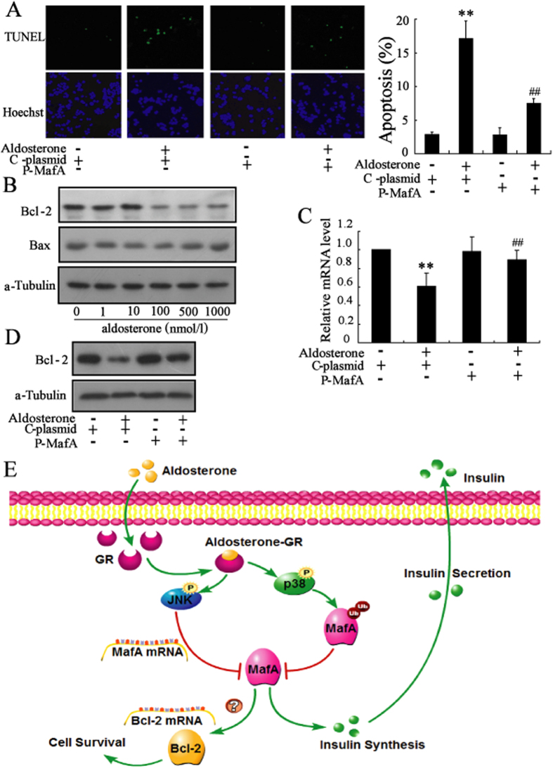
Figure 7. Up-regulation of MafA expression protects β-cells from aldosterone-induced apoptosis.
(A) After transfection with the MafA over-expression plasmid (P-MafA) or control plasmid (C-plasmid) for 24 h, Min6 cells were treated with aldosterone (100 nmol/L) for an additional 72 h. Transfection with P-MafA reversed the aldosterone-induced apoptosis of Min6 cells. (B) Aldosterone significantly decreased the protein level of Bcl-2 in a dose-dependent manner. (C) Transfection with P-MafA in Min6 cells reversed the aldosterone-induced decrease of Bcl-2 mRNA level. (D) Transfection with P-MafA in Min6 cells reversed the aldosterone-induced decrease of Bcl-2 protein level. (E) Diagram depicting the mechanism of aldosterone-induced β-cell failure. **P < 0.01, compared to control. ##P < 0.01, compared to C-plasmid combined with aldosterone-treated group.

