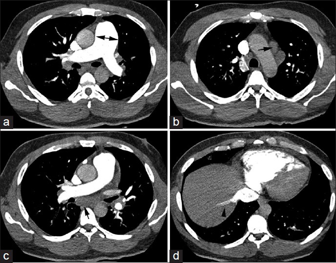
Figure 3.

42-year-old previously healthy man presented to the hospital with 6 weeks of progressively worsening exertional dyspnea and non-productive cough diagnosed with pulmonary tumor thrombotic microangiopathy (PTTM) secondary to gastric adenocarcinoma. CT chest, soft tissue window, axial slices in all panels, (a) shows an enlarged pulmonary artery (double-headed arrow), (b and c) reveal mediastinal lymphadenopathy (arrows), (d) shows reflux of contrast into the inferior vena cava (arrowhead).
