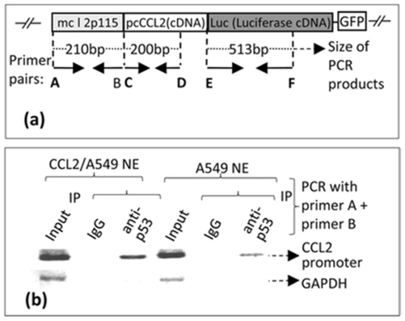
Fig. 5.

Confirmation of a stable cell line integrated with a cloned DNA (mcl2p115/pcCCL2/Luc/GFP). a. A. Schematic diagram of the cloned DNA (mcl2p115/pcCCL2/Luc/GFP) integrated into the chromosome of A549 cells (Named CCL2/A549). The arrows indicate the location and size of primers (Named primers A–F) used in PCR amplification of coding regions. These primer pairs were used for ChIP as described (Panels B). b. ChIP-detection of protein-DNA interaction between p53 and CCL2 5′UTR&promoter. The PCR-amplified specific DNA fragments are indicated with arrows.
