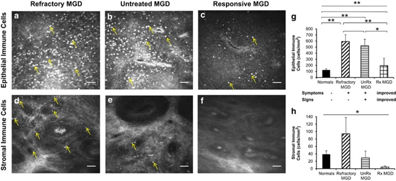
Figure 2.
Comparison of refractory MGD, untreated symptomatic MGD, and treatment-responsive MGD patients on in vivo confocal microscopy. En face in vivo confocal micrographs (HRT 3/RCM, Heidelberg Engineering, Germany) of the palpebral conjunctiva showed increased infiltration of immune cells in both the conjunctival epithelium (a–c, g; analysis of variance (ANOVA), P<0.0001) and stroma (d–f, h; ANOVA, P=0.03) of eyes with MGD-associated refractory dry eye symptoms. Epithelial immune cell density (EIC) in refractory patients (592.6±110.1 cells/mm2; a, g) was nearly five-fold greater than normals (EIC=123.7±19.2 cells/mm2 P<0.01; g), comparable to epithelial inflammation in untreated, highly symptomatic MGD patients (522.6±104.7 cells/mm2, P=0.69; b, g), and three-fold higher than that of treatment-responsive and less symptomatic MGD patients (194.9±119.4 cells/mm2, P<0.01; c, g). Stromal immune cell density (SIC) was increased several-fold in refractory patients (93.9±44.2 cells/mm2; d, h) as compared with untreated, symptomatic MGD patients (29.2±18.4 cells/mm2, P=0.33; e, h), and treatment-responsive, less symptomatic MGD patients (4.6±3.1 cells/mm2, P=0.18; f, h). Results are reported as mean±SEM. A probability value (P) of less than 0.05 was considered statistically significant (*), whereas P of less than 0.01 was considered highly statistically significant (**). Numbers in parentheses represent the number of patients per group: normals (11), refractory MGD (5), unRx MGD (3), Rx MGD (3). Axes: untreated (UnRx), treatment-responsive (Rx).

