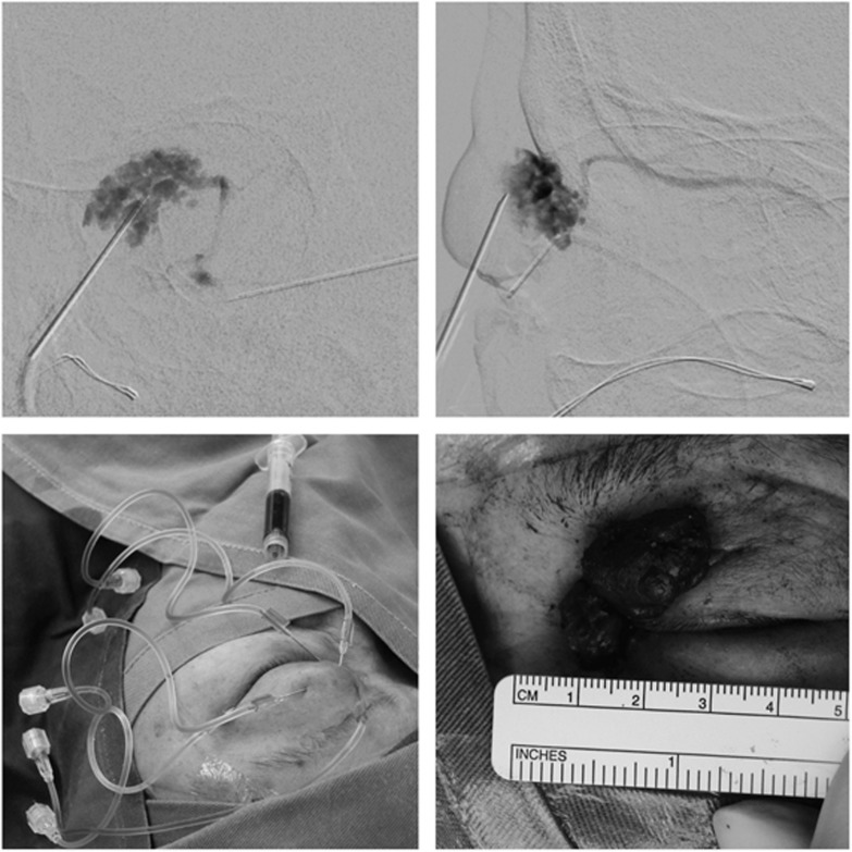
Figure 2.
Clinical photos of Case 1. (Top left) Injection of tissue glue and contrast mixture was visualized and localized with the anterior-posterior view under the biplanar digital subtraction angiographic system. (Top right) The corresponding lateral view under the biplanar angiographic system. (Bottom left) Injection of tissue glue and contrast mixture at various sites because of the multi-loculated nature of the venous malformation. (Bottom right) The glued venous malformation was excised.

