Figure 5.
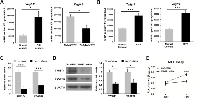
Enrichment of Vegfr2 in OIR retinas and laser CNV membrane; TWIST1 suppression with siRNA reduce VEGFR2 levels in human retinal microvascular endothelial cells (HRMECs) and its proliferation. (A) Vegfr2 mRNA expression level is increased (~3-fold) significantly in laser capture microdissected (LCM) pathological NV tufts from OIR retinas compared with normal vessels isolated from normoxic retinas at P17. Vegfr2 mRNA level was also significantly suppressed (~50%) in Tie2-Twist1cKO retinas compared with Twist1flox/flox controls at P17, *P ≤ 0.05. (B) RT-qPCR analysis also revealed a consistent enrichment of Twist1 (~2-fold) and Vegfr2 (~2-fold) mRNA expression levels in laser-induced CNV membrane compared with that of choroid without laser photocoagulation (***P ≤ 0.001). (C) Confirmation of substantial inhibition of TWIST1 mRNA expression by TWIST1 siRNA treatment. HRMECs were transfected with TWIST1 siRNA or control negative siRNA, followed by RT-qPCR analysis. VEGFR2 mRNA expression was also significantly suppressed in HRMECs transfected with TWIST1 siRNA compared with that of control group (***P ≤ 0.001). (D) Protein levels of TWIST1 and VEGFR2 were significantly downregulated (~2-fold) in HRMECs treated by TWIST1 siRNA compared with those of negative control siRNA-treated HRMECs at 48 hours (*P ≤ 0.05). (E) HRMECs were transfected with TWIST1 siRNA or control negative siRNA. At 48 and 72 hours after siRNA transfection, cell proliferation was analyzed with MTT assay. Cell proliferation in the TWIST1 siRNA-treated group was significantly lower compared with that of the control siRNA-treated group at 72 hours. MTT assay results were expressed as spectrophotometric absorbance with a detection wavelength of 540 nm at 48 and 72 hours after siRNA transfection (n = 10–11, ***P ≤ 0.001).

