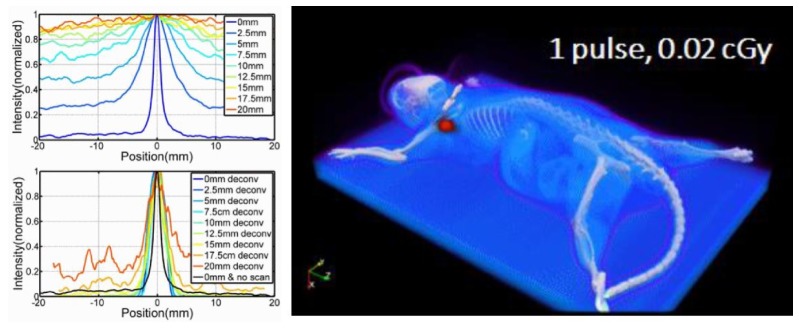
Fig. 6.

Čerenkov excited luminescence scanned imaging is demonstrated in tissue phantoms, with line-scan data from a luminescent line source in varying depths of Intralipid shown in (a) and (b) with diffuse illumination in (a) and scanned CELSI imaging in (b) illustrating the improvement in spatial resolution. The raster line scan of a mouse with luminescence in one lymph node is shown in (c), with a total body dose of 0.02 cGy [22].
