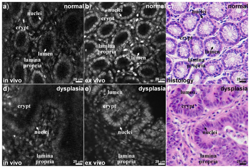
Fig. 6.
Imaging results. a) Single frame from multiphoton excited fluorescence video (Visualization 1 (13.3MB, MPG) ) of normal colonic mucosa collected in vivo at 5 frames/sec. b) Ex vivo image from normal averaged over 5 frames. c) Corresponding histology (H&E) of normal colon. Single frame from video of dysplastic crypts from colon of CPC;Apc mouse collected d) in vivo (Visualization 2 (13.3MB, MPG) ) at 5 frames/sec and e) ex vivo (averaged over 5 frames). f) Corresponding histology (H&E) of dysplasia.

