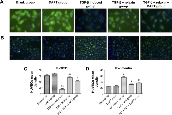
Figure 6.
Immunofluorescence staining of CD31 and vimentin to evaluate the EndMT in HUVECs.
Notes: (A) Green fluorescence represented CD31 (magnification ×400). (B) Green fluorescence represents vimentin. Blue fluorescence represented the nucleus of the cells (magnification ×200). All experiments were performed in three repetitions. (C) and (D) show the mean density of CD31 and vimentin, respectively. Data are mean ± SEM. *P<0.05, **P<0.01 vs control, #P<0.05, ##P<0.01 vs TGF-β, £P<0.05 vs TGF-β + RLX (200 ng·mL−1).
Abbreviations: EndMT, endothelial–mesenchymal transition; HUVECs, human umbilical vein endothelial cells; TGF-β, transforming growth factor β; RLX, relaxin; DAPT, N-[N-(3,5-difluorophenacetyl)-1-alanyl]-S-Phenylglycine t-butyl ester; SEM, standard error of the mean.

