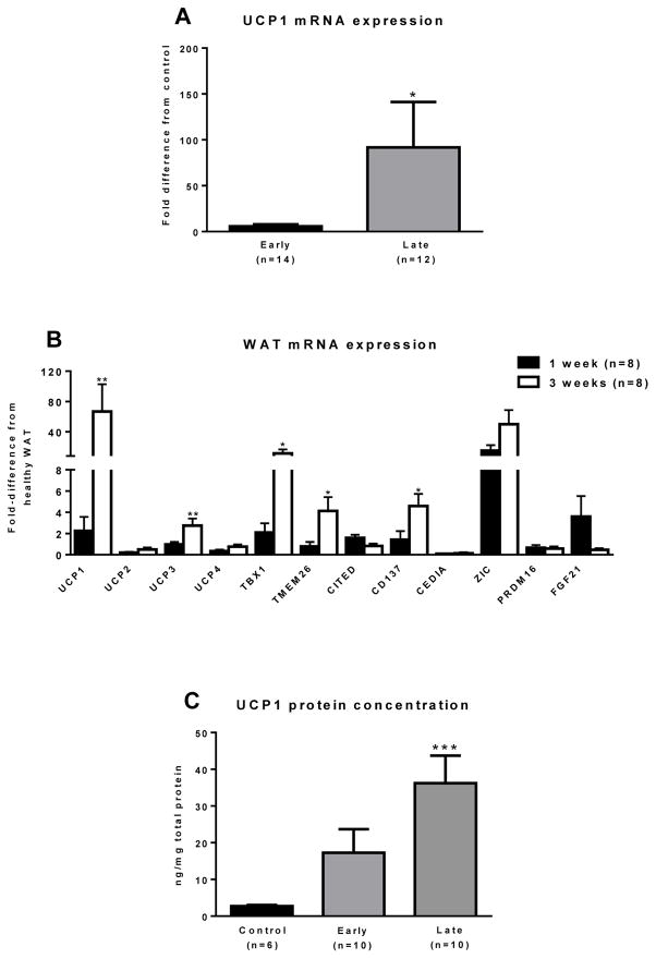
Figure 2. Molecular evidence of browning in sWAT.
(A) UCP1 mRNA expression was determined in sWAT from 9 healthy controls, 14 burned children in the early burn group and 12 burned children in the late burn group. Controls had minimal UCP1 expression. UCP1 expression was ~15-fold greater in the late group vs. the early group and ~80-fold greater in the late burn group vs. the healthy control group (*, P<0.05 vs. healthy control group). (B) UCP1 and its homologs UCP2-4 were determined in sWAT. There was a ~ 75-fold increase in sWAT UCP1 following burn trauma (P<0.01). Similarly, UCP3 was also significantly increased in sWAT following burn (P<0.01). UCP2 and UCP4 were not significantly altered by burn. The beige adipocyte markers TBX1, TMEM and CD137 were all significantly elevated in the sWAT of burn victims at the late time point. The brown adipocyte marker Zic1 was greater in sWAT of burn victims vs. controls, in particularly, Zic1 mRNA expression was an ~ 50-fold greater in burn patients sWAT at the late time point vs. controls (P<0.01). All data are presented as fold-change from healthy sWAT (*, P<0.05 and **, P<0.01 vs. healthy sWAT, respectively). (C) UCP1 protein concentration, as determined by ELISA, was significantly higher in the sWAT samples from the late burn group vs. the healthy control group (***, P<0.001 vs. healthy control group).

