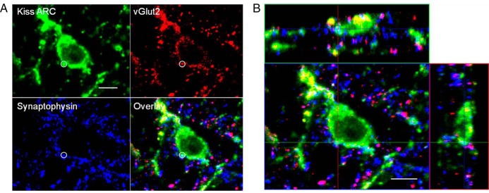
Figure 3.
A, Confocal 1-μm-thick optical section through an ARC kisspeptin cell from a prenatal T ewe, showing a triple-labeled kisspeptin/vGlut2/Syn-positive input (white circle). B, Orthogonal views from the same Z-stack, showing the direct contact between this terminal and the ARC kisspeptin neuron in all 3 planes. Scale bar, 10 μm.

