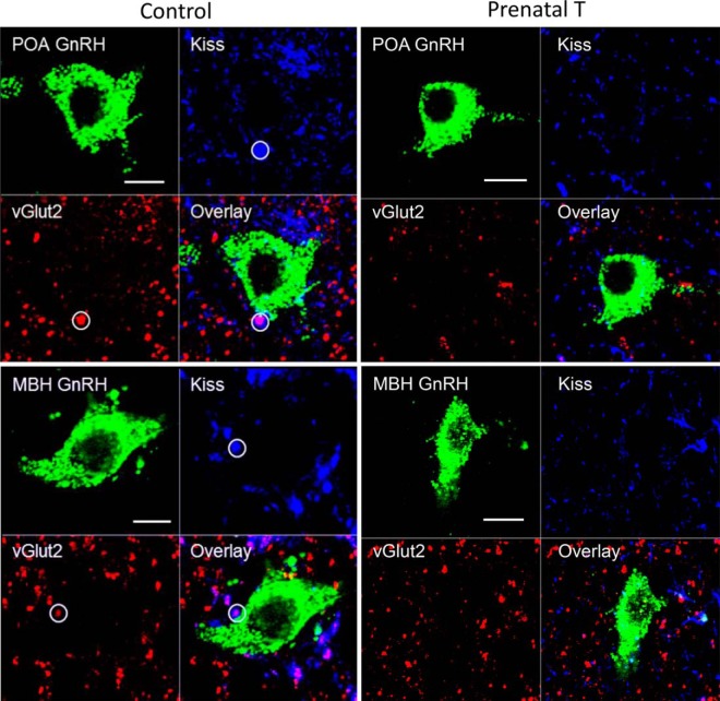
Figure 6.
Confocal 1-μm-thick optical sections through GnRH neurons in the POA (top panels) and MBH (bottom panels) from control (right) and prenatal T (right) ewes, showing examples of inputs in control ewes (white circles) that were double-labeled for kisspeptin (blue) and vGlut2 (red), along with the overlay of the 3 channels. Scale bar, 10 μm.

