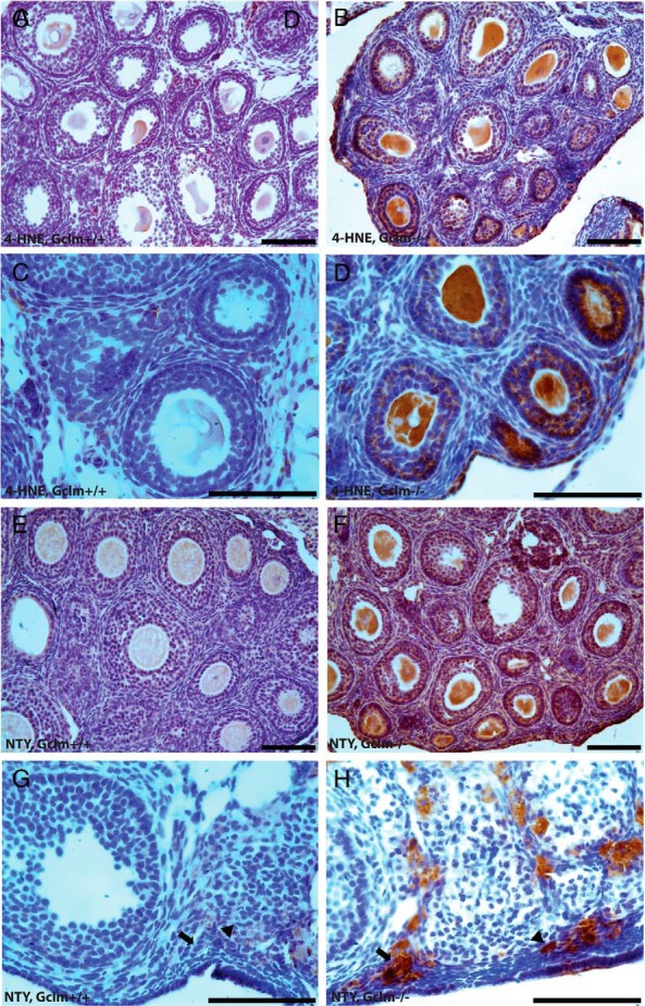
Figure 3.

Ovarian oxidative damage in Gclm−/− mice. Ovarian oxidative damage in granulosa and theca cells was scored for immunostaining with oxidative damage markers as described in Materials and Methods. Representative images of immunostaining with each oxidative damage marker in the granulosa and theca cells are as follows: lipid peroxidation (4-HNE; A–D) and protein NTY (E–H) in the ovaries of 21-day- and 9-month-old Gclm+/+ and Gclm−/− mice. A, Representative image of immunostaining for 4-HNE in the ovary of a 21-day-old Gclm+/+ mouse. B, Representative image of immunostaining for 4-HNE in the ovary of a 21-day-old Gclm−/− mouse. C, Representative image of immunostaining for 4-HNE in the ovary of a 21-day-old Gclm+/+ mouse. D, Representative image of immunostaining for 4-HNE in the ovary of a 21-day-old Gclm−/− mouse. E, Representative image of immunostaining for NTY in the ovary of a 21-day-old Gclm+/+ mouse. F, Representative image of immuonstaining for NTY in the ovary of a 21-day-old Gclm−/− mouse. G, Representative image of immunostaining for NTY in the ovary of a 9-month-old Gclm+/+ mouse. The black arrows indicate primary follicles and the black arrowheads indicate primordial follicles. H, Representative image of immunostaining for NTY in the ovary of a 9-month-old Gclm−/− mouse. All scale bars, 100 μm.
