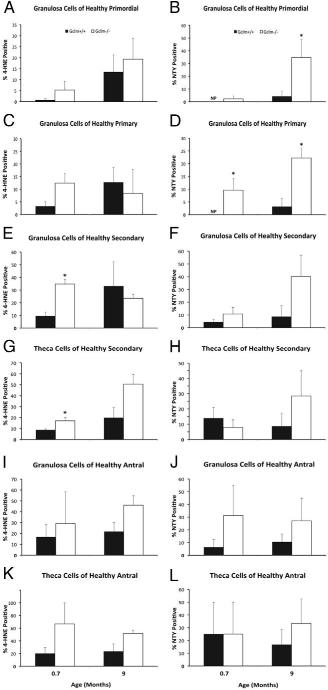
Figure 4.

Increased ovarian oxidative damage in Gclm−/− mice. Ovarian oxidative damage in granulosa and theca cells of 21-day (0.7 months) and 9-month-old mice was scored for immunostaining with oxidative damage markers as described in Materials and Methods. Quantification of immunostaining with each oxidative damage marker in granulosa cells of healthy primordial follicles are as follows: A, lipid peroxidation (4-HNE); B, protein NTY. C and D, Quantification of immunostaining with each oxidative damage marker in granulosa cells of healthy primary follicles as follows: C, 4-HNE; D, NTY. E and F, Quantification of immunostaining with each oxidative damage marker in granulosa cells of healthy secondary follicles as follows: E, 4-HNE; F, NTY. G and H, Quantification of immunostaining with each oxidative damage marker in theca cells of healthy secondary follicles as follows: G, 4-HNE; H, NTY. I and J, Quantification of immunostaining with each oxidative damage marker in granulosa cells of healthy antral follicles as follows: I, 4-HNE; J, NTY. K and L, Quantification of immunostaining with each oxidative damage marker in theca cells of healthy antral follicles as follows: K, 4-HNE; L, NTY. NP, no follicles scored positive for the given marker. Data presented are means ± SEM of the percentage of follicles with positive staining for the indicated marker from three or five experiments. Statistical significance was analyzed using an independent-samples t test after arcsine square root transformation and nonparametric Mann-Whitney U test; the results of both tests were similar. *, Significant difference between genotypes at P < .05 (n = 4–6/group).
