Figure 5.
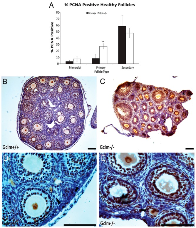
Increased granulosa cell proliferation in Gclm−/− mouse ovaries. Granulosa cell proliferation was scored as immunostaining with PCNA as described in Materials and Methods. A, Quantification of immunostaining with PCNA in granulosa cells of healthy primordial, primary, and secondary follicles in 21-day-old female mice. *, Significant difference with genotype at P < .05 (n = 4–6/group). B, Representative image of immunostaining with PCNA in the ovary of 21-day-old Gclm+/+ mice. C, Representative image of PCNA immunostaining in the ovary of 21-day-old Gclm−/− mice. D, Representative image of PCNA immunostaining in the ovary of 21-day-old Gclm+/+ mice. E, Representative image of PCNA immunostaining in the ovary of 21-day-old Gclm−/− mice. The black arrows in D and E indicate primary follicles. Data presented are means ± SEM of the percentage of follicles with positive staining for PCNA from four experiments. Statistical significance was analyzed using an independent-samples t test after arcsine square root transformation. All scale bars, 100 μm.

