Fig. 2.
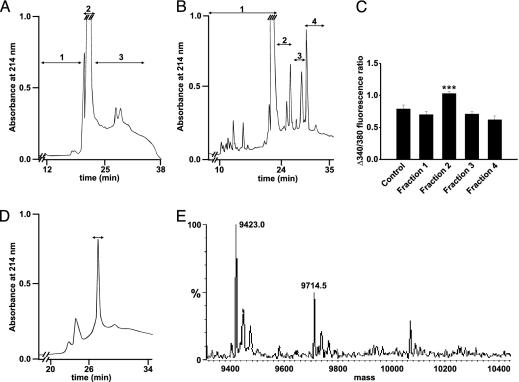
Stepwise separation and identification of the active fraction in T1D serum.(A) After the first RP-HPLC separation, the fraction marked 3 was found to give a higher increase in [Ca2+]i. (B) Fraction 3 (A) was rerun on RP-HPLC under identical conditions. The fractions were again tested for [Ca2+]i-stimulating activity (C), and one positive fraction (fraction 2) was identified. (C) Pancreatic β cells incubated with four fractions from RP-HPLC of diabetic sera from B (n = 6, 11, 12, 11, and 10, respectively). ***, P < 0.001 versus control. (D) The active fraction (B) was rechromatographed. The fraction, inducing a higher increase in [Ca2+]i when β cells were depolarized with high concentrations of K+, is marked with a bar. (E) The active fraction from C was analyzed by electrospray MS.

