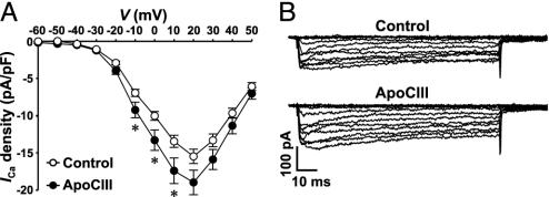
Fig. 4.
Interaction of apoCIII with the voltage-gated L-type Ca2+ channel. (A) Summary graph of current density–voltage relationships. ApoCIII-treated cells (filled circles, n = 56) and control cells (open circles, n = 55) were depolarized to potentials between -60 and 50 mV, in 10-mV increments, from a holding potential of -70 mV. *, P < 0.05. (B) Sample whole-cell Ca2+ current traces from a control cell (cell capacitance: 4.3 pF) and a cell incubated with apoCIII (cell capacitance: 4.2 pF). Cells were depolarized by a set of voltage pulses (100 ms, 0.5 Hz) between -60 and 50 mV, in 10-mV increments, from a holding potential of -70 mV.

