
Fig. 1.
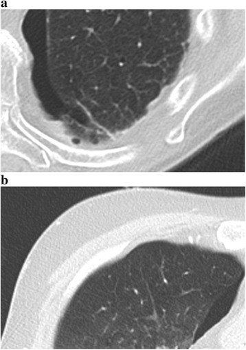
Patient versus Thiel cadaver chest CT image. Normal lung parenchyma illustrating nodular hypodense structures in a low density area, nodular hyperdense structures in a low density area, inter- or intralobular septa and the visceral pleura in both a patient (a) and a Thiel cadaver (b) chest CT image