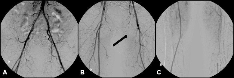
Fig. 1.

Lower extremity angiography (2004). a Both common iliac arteries showed patent flow without stenosis or occlusion. b The left superficial femoral artery was occluded at its origin (arrow). c The left distal superficial femoral artery was reconstituted by an abnormal corkscrew collateral blood flow from the left deep femoral artery
