Fig. 2.

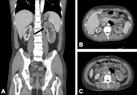
Contrast-enhanced abdominal computed tomography. a Coronal and (b, c) transverse scans showed left kidney enlargement with a multifocal infarcted area (arrows). Neither renal artery was traced from the proximal part on computed tomography

Contrast-enhanced abdominal computed tomography. a Coronal and (b, c) transverse scans showed left kidney enlargement with a multifocal infarcted area (arrows). Neither renal artery was traced from the proximal part on computed tomography