Figure 5.
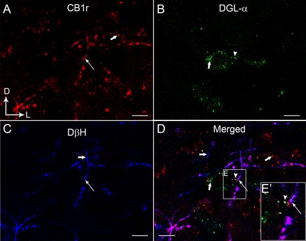
Confocal fluorescence micrographs showing cannabinoid receptor type 1 (CB1r, A), 1,2 diacylglycerol lipase-α (DGL-α, B) and dopamine-β-hydroxylase (DβH, C) in the rat frontal cortex (FC). CB1r was detected using a rhodamine isothiocyanate-conjugated secondary antibody (TRITC donkey anti-guinea pig; red) and DGL-α was detected using a fluorescein isothiocyanate-conjugated secondary antibody (FITC-donkey anti-rabbit; green). DβH-labeling was detected using Cy5 donkey anti-mouse secondary antibody (blue). CB1r and DβH appeared punctate throughout. The merged image (D) shows co-localization of CB1r and DβH in the same processes (white thin arrows) and in close proximity to DGL-α (arrowhead) in the FC. Thick white arrows indicate single-labeling (CB1r, DGL or DβH). The inset shows a higher magnification view of the area outlined by the boxed region showing close associations between DβH/CB1r and DGL-α. Scale bars, 100 μm.

