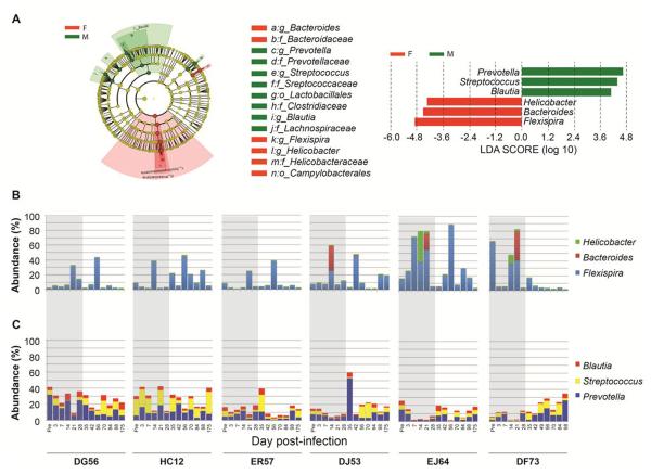
Figure 5. Gender specific microbial communities in infected animals.
Bacterial taxa informative of differences in mucosal bacterial communities between infected male and female RMs were identified by LEfSe. Differential representation of bacterial lineages, as highlighted by small circles and by shading, between male (DG56, HC12, ER57) and female (DJ53, EJ64, DF73) RMs during acute stage (d3 - 28) is mapped to a taxonomic cladogram constructed with known bacterial taxa (A). The circles represent phylogenetic levels from phylum to genus inside out. Diameter of each circle is proportional to taxon abundance; red, females; green, males. Histogram of bacterial genera enriched (LDA scores >4) during acute infection is also shown; red, female RMs; green, male RMs. Histograms showing temporal changes in relative abundance of genera significantly enriched in female and male rhesus macaques during the course of infection (B and C). Flexispira (11.69% in females vs. 3.42% in males), Bacteroides (0.37% vs. 0.09%), and Helicobacter (0.63% vs. 0.11%) were significantly enriched in female mucosal samples (B), while Prevotella (4.10% vs. 10.87%), Streptococcus (0.80% vs.2.20%), and Blautia (1.52% vs. 3.78%) were overly represented in male mucosal samples (C). Shaded area demarks the pre- and acute phase of infection.

