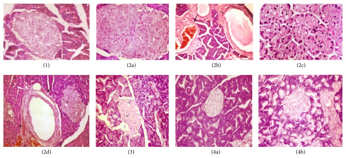
Figure 3.
Pancreas of control group (1) showing no histopathological changes. Sections ((2a)–(2d)): pancreas of hyperglycemic rats showing β-cells hyperplasia of Langerhans islets (2a), dilatation of pancreatic duct (small arrow) and congestion of blood vessel (large arrow) (b), vacuolar degeneration of epithelial lining pancreatic acini associated with pyknosis of their nuclei (2c), and vacuolation of sporadic of β-cells of Langerhans islets (small arrow), cystic dilatation of pancreatic duct (large arrow), and congestion of periductal blood vessel (arrow head) (2d). Section (3): pancreas of hyperglycemic + G. asiatica fruit extract (100 mg/kg) group showing vacuolation β-cells of Langerhans islets. Sections ((4a) and (4b)): pancreas of hyperglycemic + G. asiatica fruit extract (200 mg/kg) group showing no apparent histopathological changes (H&E ×400).

