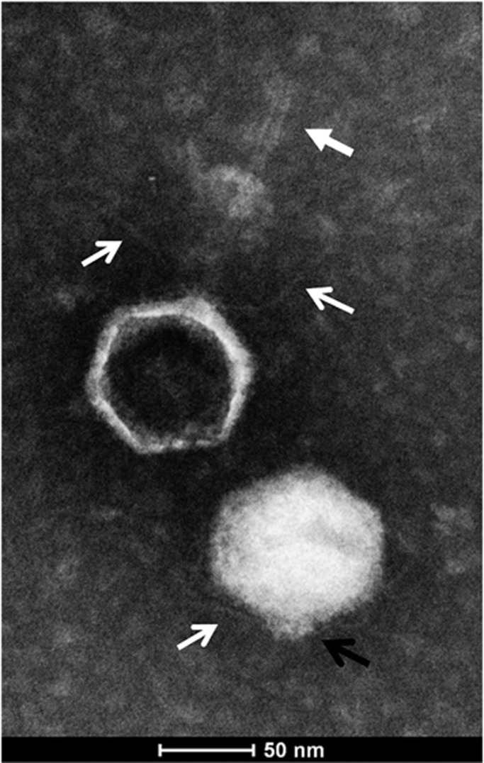
Figure 1.
General features of cyanophage S-EIV1. Transmission electron micrograph of S-EIV1 negatively stained with 2% phosphotungstic acid reveals icosahedral capsids with fine tail fibers (open white arrows) and short spiky extensions (black open arrow) that were present in filled and empty capsids, whereas delicate tail-like structures were only associated with empty capsids (closed white arrow).

