Fig. 3.

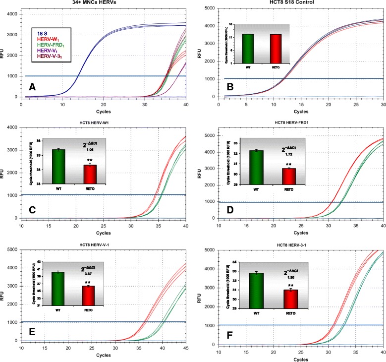
Differential expression of HERVs in HCT8WT/RETO, analyzed by real-time PCR. a: Amplification pattern of different HERVs in CD34+ mononuclear cells (MNC) from umbilical blood. The cells express all analyzed HERV variants. b: 18S endogenous control. c: Differential expression of HERV-WE1 (2−ΔΔCt 1.06). d: Degree of difference of HERV-FRD1 (2−ΔΔCt 1.72). Degree of difference of HERV-V1 (e, 2−ΔΔCt 3.87) and HERV 31 (f, 2−ΔΔCt 1.80) transcripts in HCT8 WT/RETO. In HCT8RETO, all HERV are overexpressed compared to HCT8WT. Red, WT; green, RETO, n = 3 independent experiments
