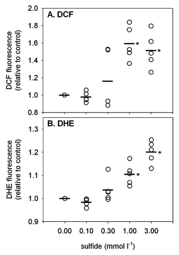
Figure 1.
Fluorescence intensity of 2′,7′-dichlorofluorescein (DCF; A) and dihydroethidine (DHE; B) in Glycera dibranchiata coelomocytes exposed to sulfide in vitro. Fluorescence is presented relative to the mean of the control samples (0 sulfide) for each worm, with circles representing individual data and horizontal lines representing the means for each treatment (coelomocytes from five worms). Asterisks indicate significant difference in the mean compared to the control.

