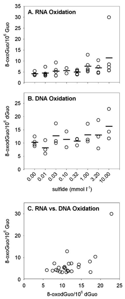
Figure 5.
A, B, Oxidative damage to RNA nucleosides (8-oxoGuo, A) and DNA nucleosides (8-oxodGuo, B) in body wall of Glycera dibranchiata coelomocytes exposed to sulfide in vivo. Concentrations of oxidized nucleosides are presented per 106 undamaged nucleosides, with circles representing individual data and horizontal lines representing the means for each treatment (three worms per treatment for RNA, two or three worms per treatment for DNA). Note that although the data in A and B were analyzed by linear regression, the abscissas in the plots are log scaled. C, Ratio of oxidatively damaged nucleosides from RNA and DNA. 8-oxoGuo = 8-oxo-7,8-dihydroguanosine; 8-oxodGuo = 8-oxo-7,8-dihydro-2′-deoxyguanosine; Guo = guanosine; dGuo = deoxyguanosine.

