
Fig 4. ConA-induced T and NKT lymphocyte activation is reduced in iron deficiency.
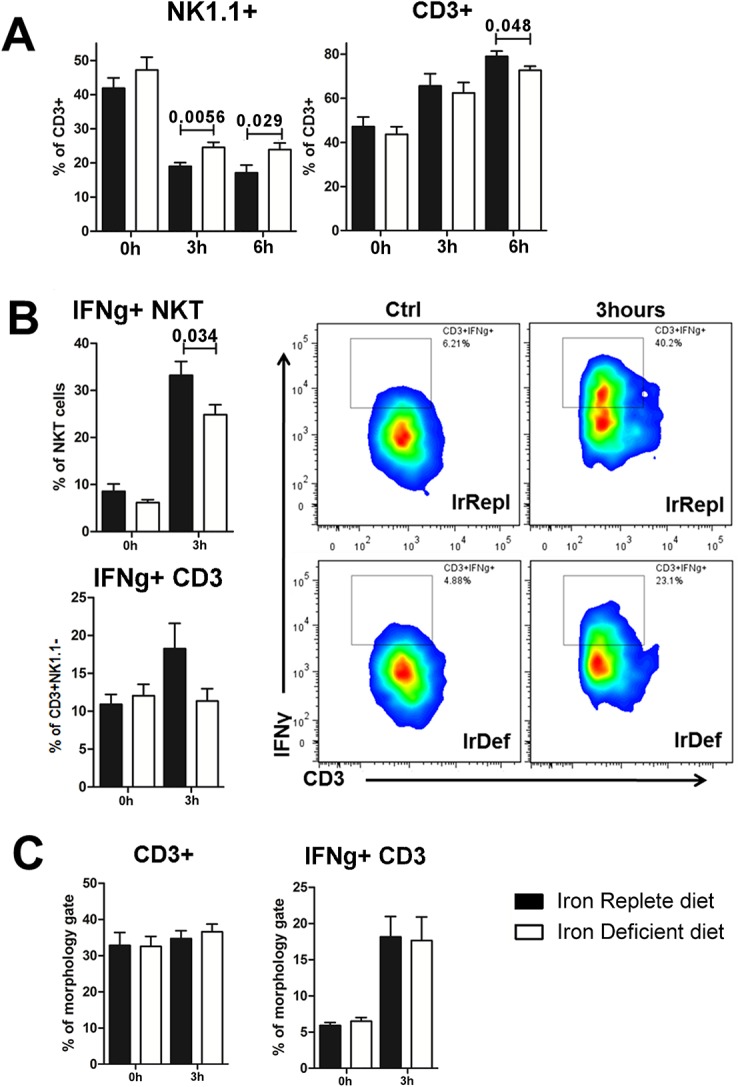
(A) Intra-hepatic frequency of CD3+ expressing NK1.1 and CD3+NK1.1- at baseline, 3 hours and 6 hours after ConA injection. (B) Intra-hepatic frequency of IFNγ production by NKT cells and T cells, defined as CD3+NK1.1+ and CD3+NK1.1-, respectively, (left panel) and representative staining of IFNγ production by NKT cells (right panel) (C) Frequency of splenic CD3+ NK1.1- T cells and the percentage of these ones producing IFNγ.