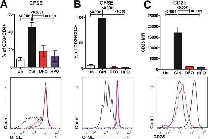
Fig 5. Iron chelation impairs CD4+ T cell activation and proliferation in vitro.
(A) Percentages of CD3+CD4+ cells from splenocytes undergoing in at least one division are represented with representative fluorescent histogram in the lower part. Splenocytes were incubated 96 hours in presence of ConA (0.1mg/mL) and in presence of low doses of iron chelator (HPO CP182, 5μM or DFO, 10μM). (B) Percentages of CD3+CD4+ cells from isolated naive CD4+ cells undergoing in at least one division and (C) CD25 MFI are represented are displayed with representative fluorescent histogram in the lower part. Isolated CD4+ naïve T cells incubated 5 days in presence of anti-CD3/CD28 plate-bound antibody (2μg each). Un means unstimulated cells.

