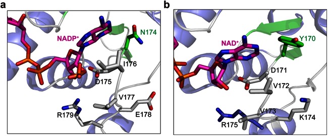
FIG 5.
Comparison between the structures of DLDH744 modeled with NADP+ and D-LDH from Aquifex aeolicus in complex with NAD+. (a) Crystal structure of DLDH744 modeled with NADP+ in the cofactor binding site. The secondary structures are colored in blue for α-helix and green for β-strand; NAD+ is colored in magenta and shown in sticks. Asn174 and other amino acids near the adenine ribose are shown in sticks. (b) The crystal structure of D-LDH from A. aeolicus bound with NAD+ was shown in same color scheme as DLDH744. Tyr170 and corresponding amino acids in D-LDH from A. aeolicus are shown in sticks.

