Fig. 5.
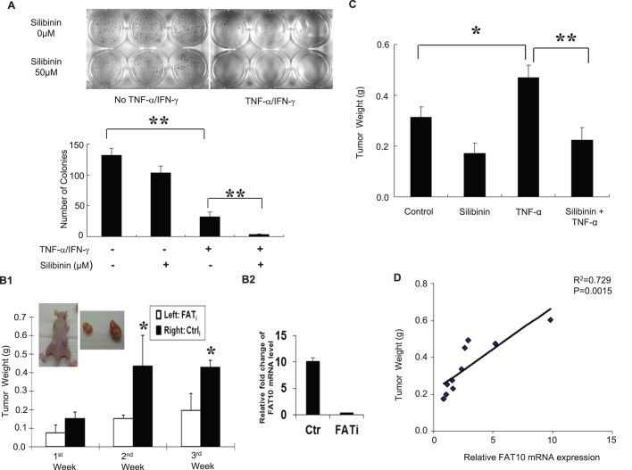
Silibinin inhibits TNF-α-induced colony formation and tumor growth. (A) Silibinin attenuates soft-agar colony formation of cells treated with TNF-α/IFN-γ (TI). Approximately 5000 HCT116 cells were seeded in soft agar and grown with TI or silibinin/TNF-α/IFN-γ (STI). Three weeks later, the colonies were stained for visualization. (B1) Inhibiting FAT10 expression with shRNA against FAT10 attenuates tumor growth. 5 μg/kg of TNF-α was administered intratumorally once after control (Ctrli) or FAT10 (FATi) shRNA containing HCT116 cells were inoculated subcutaneously into 6-week-old male athymic nude mice. Four weeks after injection, mice were sacrificed and the weights of their primary tumors were measured. (B2) Comparison of FAT10 mRNA level in FATi stable cells or control HCT116 cells injected to the mice. (C) Silibinin inhibits tumor growth. Mice with subcutaneously injected HCT116 cells and intratumorally administered TNF-α were either gavaged with saline or 100 mg/kg silibinin 5 days a week and tumor weight was determined after 7 weeks. All data in the graphs are shown as mean±s.e. *P<0.05; **P<0.01. (D) Relationship between tumor size and FAT10 expression was measured using Pearson's method.

