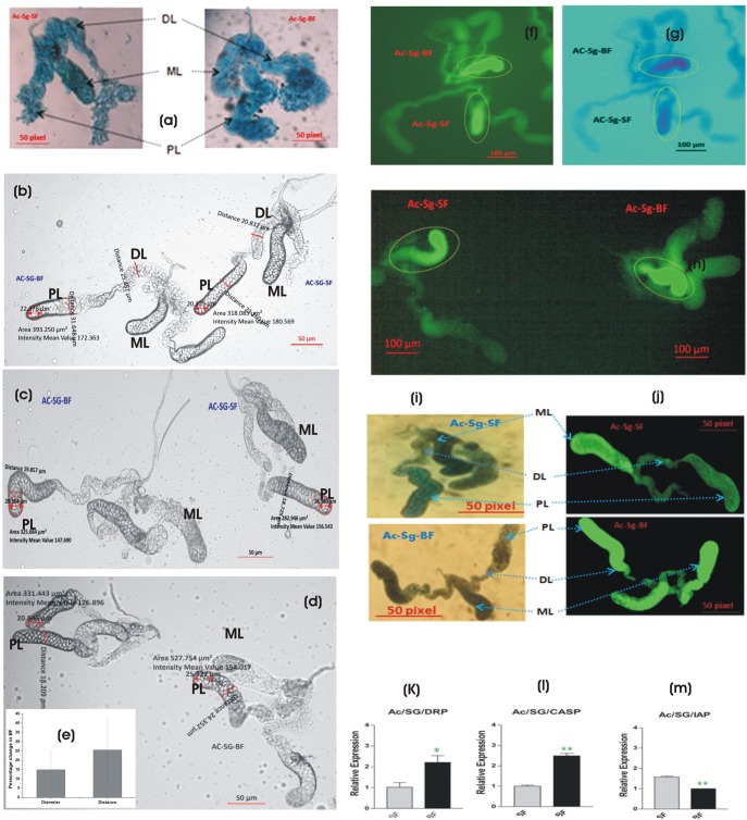
Fig. 8.
Impact of blood meal on adult female mosquito salivary glands. (A) Blood meal alters the morphological architecture, resulting in the swelling and extension of the salivary lobes. Nile blue stained glands observed under simple compound microscope (10× zoom) and captured with 7× mega pixels camera (Sony). (B-D) Phase contrast microscopy and (E) quantitative estimation of morphological features altered in response to first blood meal in the salivary glands (see text for detail). (F-J) TUNEL assay demonstrating apoptotic response in (F-H) the medial lobe (ML) (yellow circle) of the salivary glands and (I-J) in the distal and proximal lateral lobes (DL, PL) post blood meal. Loss of green color after staining with methylene blue is due to defragmentation of nuclear DNA. Note: image G is negative to image F with custom color background provided within the software for more better intensity resolution. (K-M) Relative expression of positive (DRP, draper; CASP, caspase) and negative (IAP, inhibitor of apoptosis) apoptotic marker genes in the salivary glands. Ac, Anopheles culicifacies, SG, salivary gland. (*P≤0.05; **P≤0.005). Error bar represents standard deviation from three biological replicates.

