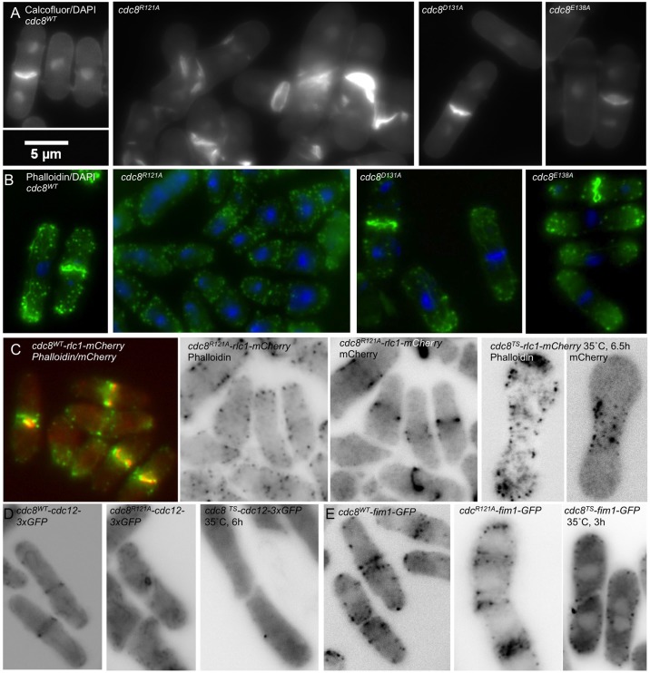
Fig. 2.
Morphology and cytoskeleton organization of cdc8wt, cdc8R121A, cdc8D131A, cdc8E138A. (A) Fixed wildtype (SH30) and mutant cells stained with Calcofluor to show septum and septal material and DAPI to show nuclei. The cdc8R121A cells (SH39) had disorganized septa and abnormal cell shapes. Cells with up to four nuclei were observed. cdc8D131A (SH43) and cdc8E138A (SH34) appeared normal. (B) Staining of the actin cytoskeleton using Alexa-phalloidin (green), and nuclei with DAPI (blue) showed dispersed patches, the absence of actin cables and poorly condensed actin rings in cdc8R121A. The other strains appeared normal. (C) cdc8wt-rlc1-mCherry (SH49) and cdc8R121A-rlc1-mCherry (SH52) cells fixed and stained with Alexa-phalloidin (green) to visualize the actin cytoskeleton. m-Cherry (red) shows localization of myosin II in the contractile ring in cdc8wt. In cdc8R121A-rlc1-mCherry myosin II assembles into contractile rings despite the absence of organized actin in contractile rings or cables. For comparison, in cdc8-27-rlc1-mCherry (SH81), a temperature-sensitive mutant, neither the actin (Alexa phalloidin) nor myosin II (rlc1-mCherry) assembled into a contractile ring at the restrictive temperature (35°C). Other cells were grown and fixed at 30°C. (D) Formin-GFP (Cdc12p-GFP) fluoresence. Formin localized in rings in wildtype (SH57) but, like Rlc1p-mCherry, formin (Cdc12p) assembled into abnormal rings in cdc8R121A (SH60) (live cells). No midline localization was observed in a temperature sensitive strain (SH87) at the restrictive temperature (fixed cells at 35°C). (E) Fimbrin-GFP fluorescence (live cells confocal image). Fim1p-GFP localized in the midline region in cdc8wt (SH77) and cdc8R121A cells (SH75) (live cells), but not in the temperature-sensitive strain (SH83) at the restrictive temperature (cells grown and fixed at 35°C). The localization of fluorescent proteins was normal in cdc8D131A and cdc8E138A strains (supplementary material Fig. S3).

